PUBLISHED IN TCI WEEKLY NEWS
May 9th 2015

When a dentist examines your mouth, they learn a lot about the health of your teeth, gums and other areas of the mouth just by looking. However, what a visual examination does not reveal is what is happening under the gums. Dental X-rays (or radiographs) are able to reveal what is happening inside and between the teeth, and within the bone and so provide the dentist with a fuller picture to faciliate an accurate diagnosis.
Valuable Diagnostic Tool
An X-ray is taken if the dentist needs to investigate an area further; they are considered a special investigation and should not be taken routinely. They are used to check for cavities and evaluate the extent of decay. And because they show the root of the tooth, the presence of any cysts, abscesses and other masses can be diagnosed. Congenitally missing or impacted teeth such as wisdom teeth are often identified this way, and the presence and extent of bone loss due to periodontal disease is easily seen through dental X-rays as well.
Types of X-rays
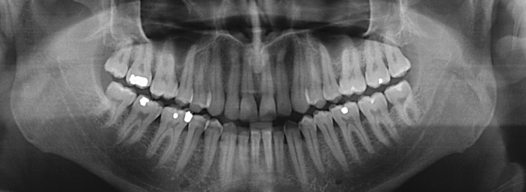
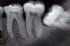
Most modern dental clinics nowadays with have digital X-rays which mean the old- fashioned concept of having to develop films is redundant.  The X-ray immediately uploads to the dentist’s computer where he can study the image. This also has the added benefit of being digitally attached to your dental records and can be emailed if ever required. There are several types of radiographs commonly used in the dental office: periapical shows one or a few teeth from the crown right down to the tips of the root, bitewing shows a row of back teeth, and panoramic which shows the whole mouth. It is common to go to a hospital for a panoramic X-ray: this is true on Provo.
Dental X-rays Safety
X-ray machines are designed to minimize radiation and as technology as improved, the length of exposure to the X-rays has continued to reduce so you can be reassured that your exposure is negligible and the processes are safe. In my clinic, the technology is very modern and the patient is only exposed to the X-rays for four hundredths of a second.  Also, both my nurse and I wear X-ray monitoring badges which measure the amount of X-ray exposure over time, to ensure we are both working safely. Some (old fashioned) protocols dictate the use of a lead apron for the patient, although the best practice I follow actually recommends against this as it can scatter the x-rays and therefore increase exposure.
Any patient must inform the dentist if they are pregnant, as X-rays for routine treatment would be postponed until after the baby is born.